A 3D Lipid Atlas of the Mouse Brain Finds 539 Chemical Zones That Shift Sharply During Pregnancy
An EPFL-led team mapped 172 membrane lipids across the whole mouse brain at 25-micrometre resolution, publishing the atlas in Nature on September 23, 2026.
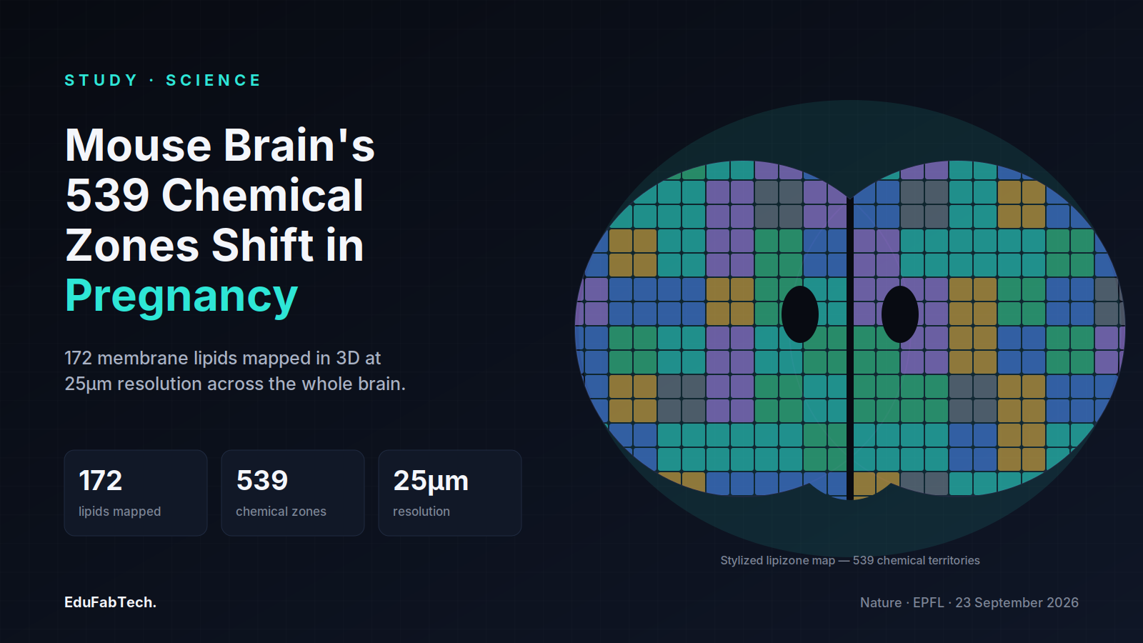
A team led by researchers at the École Polytechnique Fédérale de Lausanne (EPFL) has published the first whole-brain map of membrane lipids in the adult mouse, reporting the work in Nature on September 23, 2026. Using mass spectrometry imaging rather than the gene-expression or protein methods behind most existing brain atlases, the group led by Giovanni D'Angelo and Gioele La Manno charted 172 distinct lipid species across the brain at a spatial resolution of 25 micrometres, then grouped the resulting chemical patterns into 539 territories they call "lipizones."
What the atlas measured
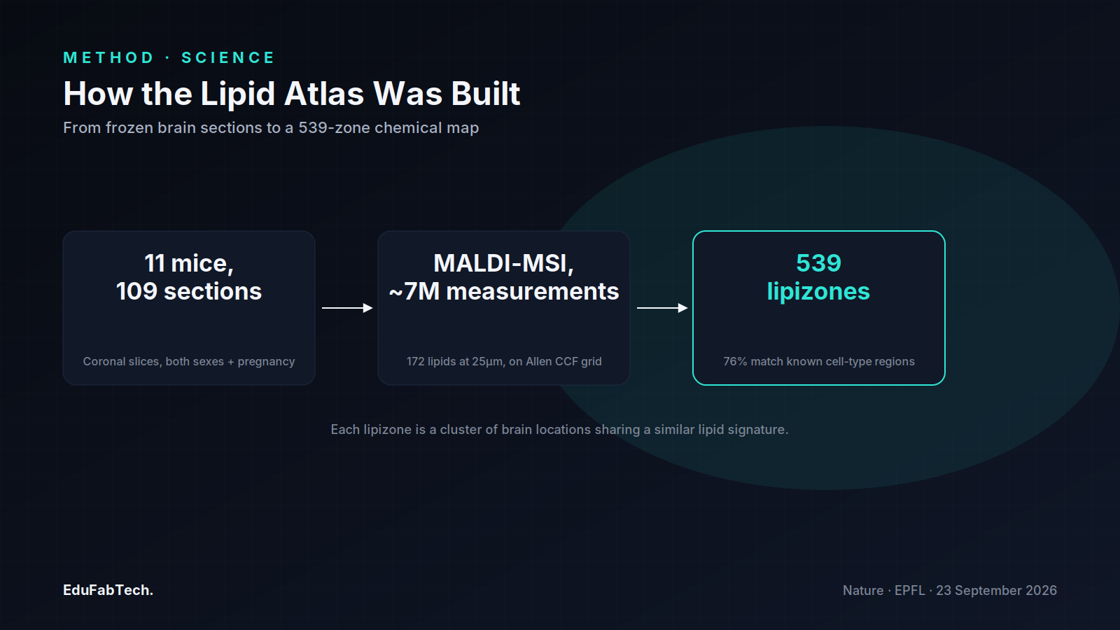
The team analyzed 109 coronal brain sections from 11 mice using matrix-assisted laser desorption/ionization mass spectrometry imaging (MALDI-MSI), producing roughly 7 million individual measurements that were then registered onto the Allen Institute's Common Coordinate Framework, the standard reference grid already used by transcriptomic and proteomic brain atlases. Each lipizone is a cluster of locations sharing a similar lipid composition, and the paper reports that 76% of lipizones significantly aligned with known cell-type-specific regions mapped by earlier single-cell studies.
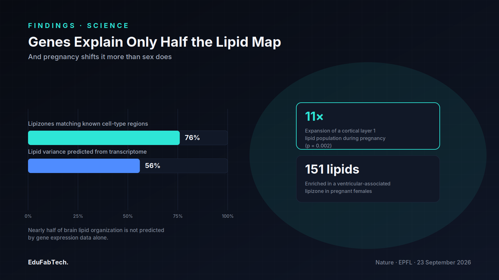
Notably, the lipid-based territories did not simply replicate maps built from gene expression. The authors found that only 56% of lipid variance could be predicted from full transcriptome data using nonlinear regression, meaning close to half of the spatial organization of brain lipids is not explained by which genes are switched on in a given location. Individual enzyme-coding genes, the paper notes, do not translate into lipid distribution in any simple one-to-one way.
A different kind of map
Existing brain atlases have mostly been built from RNA transcripts or proteins, cataloguing which genes are active in which cells. The EPFL-led group argues that lipids reveal a separate, complementary layer of organization: distinct brain regions that share a function can share a chemical "signature" of lipids even when they sit far apart and belong to different cell types. D'Angelo described the pattern as working "like postal codes, giving distant but related parts of the brain the same chemical address," according to Neuroscience News' coverage of the release. The atlas also identified a previously undescribed axis of variation among oligodendrocytes, the cells that produce the fatty myelin sheath around axons in white matter, and found biochemical zonation in the choroid plexus and ventricular walls, structures involved in producing cerebrospinal fluid.
Pregnancy reshaped the map more than sex did
The researchers also compared brains across both sexes and during pregnancy. They report that lipid changes associated with pregnancy were larger in magnitude than the baseline differences observed between males and females, with roughly 151 lipids becoming enriched in a ventricular-associated lipizone in pregnant females. Galactosylceramide, a core structural lipid of myelin, rose sharply across white matter, and a lipid-defined cell population in cortical layer 1 expanded roughly 11-fold during pregnancy (p = 0.002), part of what the authors describe as pronounced remodeling across the outer cortex. La Manno summarized the takeaway as evidence that "the brain constantly adjusts its lipids to meet the body's changing needs," per Medical Xpress's report on the paper.
Independent context
In an accompanying Nature News & Views commentary, Evan Macosko of the Stanley Center for Psychiatric Research at the Broad Institute of MIT and Harvard situates the atlas alongside the transcriptomic and proteomic maps that have dominated brain-mapping efforts in recent years, framing the lipidomic layer as a distinct axis of organization rather than a replacement for those approaches. Macosko's piece, published the same day as the primary paper, underscores that lipid zonation captures information — such as distal axon terminals and glial subtypes — that expression-based atlases can miss.
"What transfers to humans isn't the map, it's the method…"
That caveat, voiced by the paper's first author, Luca Fusar Bassini, in remarks reported by News-Medical, is central to how the authors frame their own findings. The atlas describes the mouse brain specifically; mouse and human brains differ in size, lipid composition and cellular architecture, so the anatomical positions of lipizones are not expected to carry over directly. What the authors argue does generalize is the imaging and clustering pipeline itself, which they suggest could be applied to human tissue in future work — building on smaller-scale efforts such as the 2024 lipidome atlas of the adult human brain published in Nature Communications, which sampled a more limited set of regions rather than the whole organ at cellular resolution.
Why it matters for disease research
The authors and outside commentators connect the work to a growing body of evidence that lipid dysregulation accompanies conditions ranging from depression to Alzheimer's disease and other neurodegenerative disorders. A reference map of what "normal" lipid organization looks like in a healthy brain gives researchers a baseline against which to compare tissue from disease models — for instance, to ask whether a given disorder disrupts lipizone boundaries, alters oligodendrocyte lipid signatures, or produces ventricular changes resembling those seen during pregnancy. The dataset, built on the same coordinate framework used by other major mouse brain atlases, has been made publicly available, allowing it to be layered directly against existing transcriptomic and connectivity data rather than standing as an isolated resource.
The paper is explicit that this is a first, descriptive map rather than a causal account of how or why particular lipids accumulate where they do — the authors note that understanding of lipid distribution at the subcellular level, and its functional consequences for individual cells, remains limited. Follow-up work will need to test whether specific lipizone changes are drivers of disease processes or simply markers of them.
- Luca Fusar Bassini, Gioele La Manno, Giovanni D'Angelo et al.. The lipidomic architecture of the mouse brain. Nature, 2026. doi:10.1038/s41586-026-11050-0
- Evan Macosko. Brain lipids are organized into zones — and reorganized during pregnancy. Nature (News & Views), 2026. link
- Neuroscience News staff. First 3D Brain Lipid Atlas Uncovers 539 Hidden Zones. Neuroscience News, 2026. link
- News-Medical staff. New 3D lipid atlas maps the chemistry of mouse brains. News-Medical.net, 2026. link
- Medical Xpress staff. First map of mouse brain lipids reveals over 500 biochemical zones. Medical Xpress, 2026. link